Silent (Masked) Otitis Media
 - Definition of Silent (Masked) Otitis Media - Silent Otitis Media Terminology - Chronic Silent Otitis Media - How Does Silent Middle Ear Inflammation Occur? - Eustachian Tube Problems Are Seen as The Most Common Cause of Middle Ear Fluids - Otitis Media With Effusion Does Not Necessarily Occur Following Acute Otitis Media! - Risk Factors For Silent Otitis Media - Corrected Incorrect Expression \"Middle Ear Fluid Does Not Arise From Fluid Leakage From Outside To Ear\" - How Does The Silent Otitis Media Behave? - Symptoms of Silent Otitis Media - Diagnosis of Silent Otitis Media - Treatment of Silent Otitis Media - How Can You Prevent Silent Otitis Media in Your Child? - Complications of Silent Otitis Media")
Definition of Silent (Masked) Otitis Media
Silent (masked) otitis media or silent otitis media refers to chronic pathological conditions that are clinically "undetectable" or "undetectable" behind a robust tympanic membrane (kulk membrane). If the eardrum is intact, I would like to write down the terms used synonymously or closely related to the presence of fluid behind the membrane:
• silent otitis media (SOM)
• masked middle ear inflammation
• secretory otitis media
• serous otitis media
• silent otitis media
• masked otitis media
• serous otitis media
• otitis media with effusion (OME)
Videos about "Silent Otitis Media - Masked Otitis Media - Masked Middle Ear Inflammation - Secretory Otitis Media - Serous Otitis Media - Otitis Media With Effusion (OME) - Ear Tube":
Serous Otitis Media Definition Video:
Ear Ventilation Tube (T-tube) Video:
Valsalva Maneuver Video:
Adenoid Hypertrophy Definition and Treatment Video:
Adenoidectomy Operation (Adenoid Removal) Video:
Silent Otitis Media Terminology
 |
| serous otitis media |
Chronic Silent Otitis Media
 |
| serous otitis media |
 |
| Chronic Serous Otitis Media |
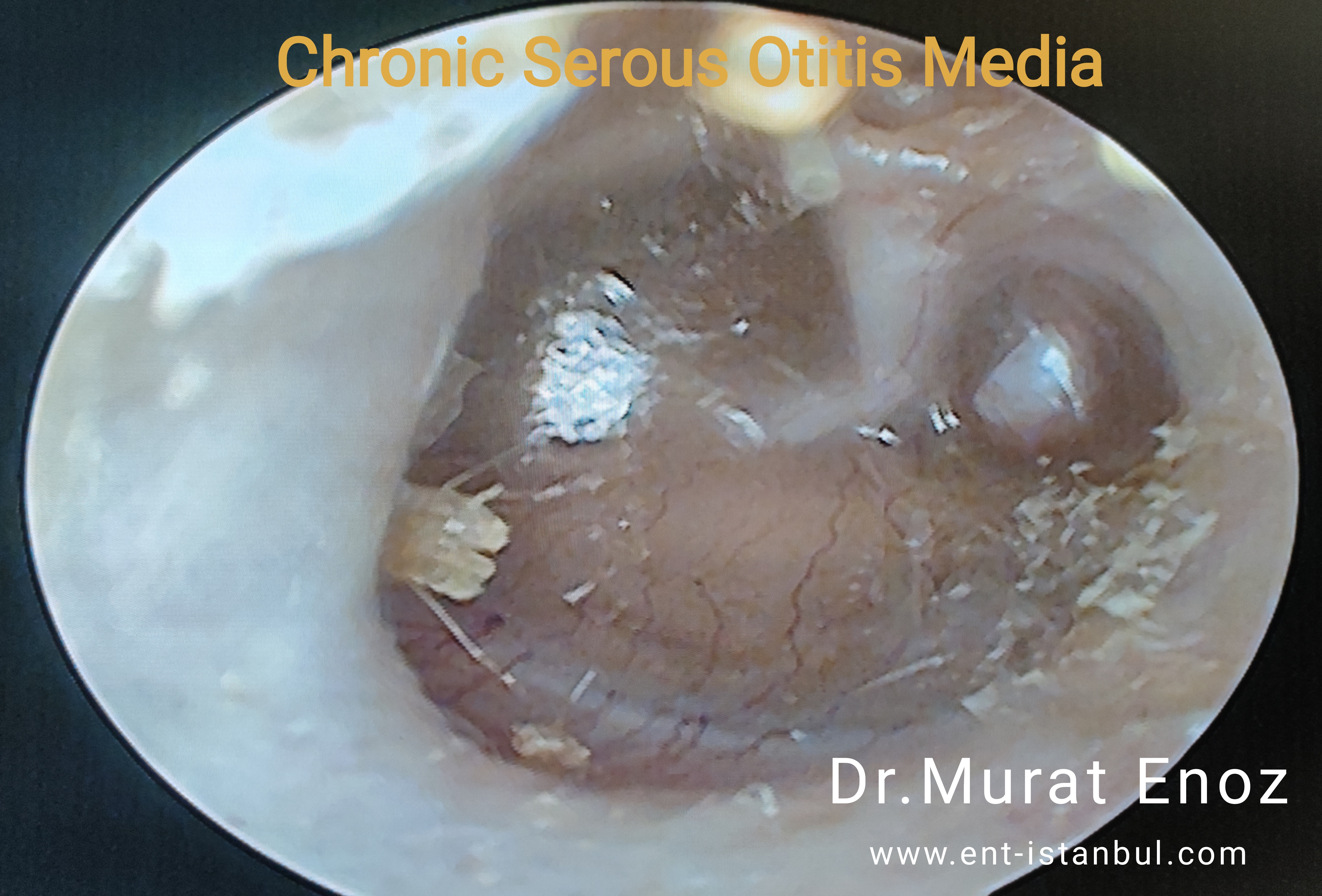
The photo above shows the eardrum taken during ear examination of a 6-year-old boy who has been sleeping with his mouth open for a long time and complaining of nasal congestion and ear congestion. it is seen that the patient has a dark viscous and brownish liquid image behind the eardrum, retraction in the eardrum and a retraction pocket in the posterior upper quadrant. Irregularities in the eardrum and refractions in light reflection can be noticed, the effects of long-term negative pressure in the middle ear caused changes in the eardrum. Ear ventilation tube insertion + adenoid removal operations are planned for the treatment of chronic effusion or serous otitis media.
How Does Silent Middle Ear Inflammation Occur?
After acute inflammation is resolved, otitis media (OME) with effusion may occur during the recovery period of acute otitis media (AOM - acute otitis). Among children with acute otitis media, 45% had persistent effusion after one month, and this number decreased to 10% after 3 months.There are two basic theories of acute otitis media:
1. The classical explanation suggests that Eustachian tube dysfunction is a necessary premise. The Eustachian tube is traditionally defined to provide 3 main functions: balancing the pressure between the middle and the outer ears, clearing the secretions and protecting the middle ear. A variety of conditions can be caused by dysfunction, anatomical obstruction (as in the case of nasal growth), allergy, upper respiratory tract infection, or inflammation secondary to trauma.
If the eustachian tube dysfunction is persistent, a negative pressure occurs in the middle ear due to the absorption and / or diffusion of nitrogen and oxygen into the mucosal cells in the middle ear. If the negative pressure is present for a sufficiently long period and of appropriate size, it derives the transudate from the mucosa and ultimately leads to serous, essentially sterile effusion accumulation. Because the Eustachian tube is dysfunctional, the effusion becomes a stable ideal environment for the growth of bacteria and acute otitis media.
2. As the new theory, it describes the inflammation of the middle ear mucosa caused by a response to bacteria already present in the middle ear. Inflammatory mediators released as a result of bacterial antigenic challenge induce upregulation of mucin genes. The production of a mushroom-rich effusion then provides a broad environment for bacterial growth and acute otitis media. There are studies showing that there are moist changes in oxidative stress in patients with otitis media with effusion. The investigators showed significant improvement not at the normal level of oxidants after the ventilation tubes were placed. However, the role of antioxidants in the treatment of otitis media with effusion has not yet been fully investigated.
Eustachian Tube Problems Are Seen as The Most Common Cause of Middle Ear Fluids
Eustachian tube dysfunction is almost universal in otitis media with effusion regardless of the cause of acute otitis media. In other evidence, the attachment of the Eustachian tube in animals always leads to the formation of a permanent middle ear effusion. After acute inflammation and bacterial infection are resolved, the failure of the middle ear clearance mechanism allows the middle ear effusion to continue. Many factors have been implicated in the failure of the cleansing mechanism, including ciliary dysfunction, for example, mucosal edema; hyperviscosity of effusion; and possibly a negative pressure gradient.Otitis Media With Effusion Does Not Necessarily Occur Following Acute Otitis Media!
It may also occur as a silent middle ear infection alone. theories describing the development of middle ear effusion include the release of fluid from the inflamed middle ear mucosa. This theory suggests that the middle ear mucosa was previously sensitized by exposure to bacteria, and occasionally to continue antigenic stimulation from the reflux to induce effusion. Again, multiple studies have shown that the same flora bacterium is present in otitis media with effusion as in acute otitis media; these findings confirm that the idea of a "effusion is sterile", which was once believed, is false.Risk Factors For Silent Otitis Media
Environmental factors, age and dysfunction of Eustachian tube have been associated with otitis media with effusion.Environmental factors
In addition to actual pathogens, environmental factors have been strongly associated with increased prevalence of otitis media with effusion in a number of epidemiological studies. These factors include bottle feeding, supine supine feeding, having a brother with otitis media, joining the day care (crowded nursery environment is a very big risk factor for children), living in an environment with high allergen rates, low socio-economic status, living in the home (smoking next to children, facilitating the emergence of fluid in the middle ear in children!) and having a family history of otitis media.Age
Age is another prominent factor in the development of otitis media with effusion. The eustachian tube of the baby has an almost horizontal orientation (relative to the ground), and after a few years (as in adults) it is at a 45 ° angle. In addition, the size and shape of the Eustachian tube at the time of delivery is also unfavorable for the ventilation of the middle ear, unlike the adults.The frequency of silent otitis media is highest in children aged 2-4 years, and as expected with the prevalence of otitis media with effusion, there is a significant decrease in the frequency of children older than 6 years.
In adults, it is important to recognize unilateral otitis media with effusion. This entity should be considered as a nasopharyngeal mass until it is proved to be exact. In the case of long-lasting serous otitis media in a single ear (in the case of adult unilateral middle otitis media), nasopharynx cancer (nasopharynx tumor) should be considered in the differential diagnosis.
Silent middle ear inflammation is more common in young children than older children or adults due to:
• The tube is shorter, more horizontal and flat, making it easier for bacteria to enter.
• The tube is more flexible and has a smaller opening that is easier to clog
• More frequent colds are seen because the immune system takes time to recognize cold viruses in young children and the emergence of immune response
Eustachian tube dysfunction
Disruptions in the normal opening of the eustachian tube hole in the nasopharynx are also associated with the increased prevalence of otitis media with effusion. They are usually seen in a cleft palate patient and children with Down's syndrome and other disorders affecting the palate. In addition, reduced mucociliary clearance in cystic fibrosis and higher viscosity of mucus were assumed to explain the prevalence of effusion otitis media in patients with these conditions.
The prevalence of silent otitis media is increased in children with aphid growth because of the closing of the eustachian tube with a mass effect and the potential microbes reservoir of the eustachian tube.
Diet
It has been suggested that a high-fat diet is a risk factor for otitis media with effusion in children but is in the body mass index category. For this disease, which is most commonly seen in childhood, it is difficult to find that it is directly related to the diet. I have added this information because it is in new publications.
In patients with cleft palate, silent middle ear inflammation may occur!
In children with cleft palate, otitis media with effusion are everywhere. This is because simply the tensor parent palatine muscle is not properly placed in the soft palate. Therefore, the muscle cannot open the Eustachian tube on swallowing or wide mouth opening. It causes a functional blockage of the tube.
Other risk factors for silent otitis media
Early onset of cow's milk, long sucking time and ethnic origin of Asian origin may be seen as an additional low risk in infants.
In recent years, language functions are affected, and the problem of intraabdominal lesion progression in infants who have a language that may increase the incidence of otitis media studies can be published ...
Recent studies have shown that tongue function may be increased in infants with tongue ties, which has recently been affected by tongue functions, and may increase the incidence of middle ear inflammation.
Although there is no statistically significant difference between sexes in terms of incidence or prevalence, some findings suggest that men may have a slightly higher frequency.
Allergies, irritating substances in the air and respiratory infections can cause OME. Changes in air pressure can cause the Eustachian tube to close and may affect fluid flow. These may be due to travel on the plane or sipping liquid while lying down.
Corrected Incorrect Expression "Middle Ear Fluid Does Not Arise From Fluid Leakage From Outside To Ear"
Silent middle ear inflammation does not occur due to water escape from the middle ear. The eardrum acts as a barrier between the middle ear and the outer ear. The causes of fluid collection in the middle ear are summarized above.
How Does The Silent Otitis Media Behave?
Otitis media with effusion (OME) is the leading cause of hearing loss in children. This is associated with delayed language development in children under 10 years of age, and the type of hearing loss is usually the type of transmission (there is a problem with the transmission of sound waves from the middle ear to the inner ear), and the average air conduction threshold is 27.5 decibels (dB), but rarely the presence of chronic effusion otitis media sensorineural hearing loss (nerve type hearing loss). Both prostaglandins and leukotrienes are present in high concentrations in middle ear effusions and their ability to cross the round window membrane is shown. Chronic exposure to these metabolites of Arachidonic acid may cause temporary and sometimes permanent sensorineural hearing loss.
In general, the prognosis of otitis media with effusion is good. Most OME spontaneously recover without intervention, and most of them go smoothly. Currently, 5% of children without surgical treatment have persistent otitis media with effusion. Surgical intervention provides clearance of middle ear effusion in this population; The benefits of quality of life as well as speech and language development are controversial.
Following spontaneous extrusion of ear ventilator tubes (which spontaneously excretes ear plugs) for the treatment of silent otitis media (20 to 50% of patients), recurrence of otitis media with effusion is seen and potentially combined with pressure balancing tubes and, in most cases, simultaneous adenoidectomy is required.
Symptoms of Silent Otitis Media
None of the children with silent otitis media have symptoms. Silent middle ear inflammation is not the result of an infection. Many children with silent otitis media cannot express themselves as a complaint or feel sick.
Symptoms are usually mild or minor. It may vary according to the age of the child.
A common symptom of silent otitis is a hearing problem. Behavioral changes in younger children may be a sign of hearing problems. For example, children can turn on the television louder than usual. They can also pull or mix their ears.
Older children and adults with silent otitis media often describe the voice as muffled. They can feel the fluid is full of fluid.
Diagnosis of Silent Otitis Media
During a simple ear examination of a kbb physician, the following can be seen for the quiet middle ear reminder:
• Air bubbles on the surface of the eardrum
• Smooth and glossy matte eardrum
• visible fluid behind the eardrum
• There is a non-moving eardrum when you blow a small amount of air (see the valzalva maneuver video above)
Further diagnostic methods are also available. An example is the "tympanometry test". In this test, a probe is placed in the ear. How much fluid is behind the eardrum and how much membrane collapses can be measured.
During examination with a simple otoscope, microscopic ear examination or endoscopic ear examination, middle ear fluid may be seen. This way, the diagnosis can be made easily.
Below I added a few diagnostic images that I took during the examination:
1- Normal Eardrum
 |
| Normal Eardrum |
Above is a normal eardrum image. The transparent structure of the eardrum, the light triangle, the dark image of the opening mouth of the Eustachian tube and the reflection of the ossicles are seen.
2- Acute Otitis Media
 |
| Acute Otitis Media |
Above is the appearance of acute otitis media (AOM). The veins of the eardrum are increased, there is a secretion of inflammation behind the membrane.
 |
| Acute Otitis Media |
Acute otitis media (AOM ) is also seen above. Increased vascularisation of the eardrum, inflammation of the membrane behind the secretion of the image is available, and appear inflamed eardrum outward bulging been more compared to the normal eardrum.
 |
| Acute Otitis Media |
Above, in a child with the diagnosis of acute otitis media and in whom antibiotic treatment was started, it is seen that the membrane is close to normal, there is inflammation in the middle ear and serous appearance in the middle ear, air bubbles appear in the upper part of the membrane.
3 - Silent Otitis Media


Above, the treatment of acute middle ear inflammation is provided and after 10 days of treatment there is a liquid and the fluid remaining on the back of the membrane with the effect of gravity. Matte and uneven areas on the top of the membrane surface indicate the period of improvement of the previous tympanic membrane.
 - Definition of Silent (Masked) Otitis Media - Silent Otitis Media Terminology - Chronic Silent Otitis Media - How Does Silent Middle Ear Inflammation Occur? - Eustachian Tube Problems Are Seen as The Most Common Cause of Middle Ear Fluids - Otitis Media With Effusion Does Not Necessarily Occur Following Acute Otitis Media! - Risk Factors For Silent Otitis Media - Corrected Incorrect Expression \"Middle Ear Fluid Does Not Arise From Fluid Leakage From Outside To Ear\" - How Does The Silent Otitis Media Behave? - Symptoms of Silent Otitis Media - Diagnosis of Silent Otitis Media - Treatment of Silent Otitis Media - How Can You Prevent Silent Otitis Media in Your Child? - Complications of Silent Otitis Media")
 - Definition of Silent (Masked) Otitis Media - Silent Otitis Media Terminology - Chronic Silent Otitis Media - How Does Silent Middle Ear Inflammation Occur? - Eustachian Tube Problems Are Seen as The Most Common Cause of Middle Ear Fluids - Otitis Media With Effusion Does Not Necessarily Occur Following Acute Otitis Media! - Risk Factors For Silent Otitis Media - Corrected Incorrect Expression \"Middle Ear Fluid Does Not Arise From Fluid Leakage From Outside To Ear\" - How Does The Silent Otitis Media Behave? - Symptoms of Silent Otitis Media - Diagnosis of Silent Otitis Media - Treatment of Silent Otitis Media - How Can You Prevent Silent Otitis Media in Your Child? - Complications of Silent Otitis Media")
There is an appearance of serous otitis media after otitis media. The smoothness of the membrane, the presence of the light triangle, and ossicular revival indicate that there is only middle ear fluid without serious structural changes. Air bubbles are seen on the upper part.
 - Definition of Silent (Masked) Otitis Media - Silent Otitis Media Terminology - Chronic Silent Otitis Media - How Does Silent Middle Ear Inflammation Occur? - Eustachian Tube Problems Are Seen as The Most Common Cause of Middle Ear Fluids - Otitis Media With Effusion Does Not Necessarily Occur Following Acute Otitis Media! - Risk Factors For Silent Otitis Media - Corrected Incorrect Expression \"Middle Ear Fluid Does Not Arise From Fluid Leakage From Outside To Ear\" - How Does The Silent Otitis Media Behave? - Symptoms of Silent Otitis Media - Diagnosis of Silent Otitis Media - Treatment of Silent Otitis Media - How Can You Prevent Silent Otitis Media in Your Child? - Complications of Silent Otitis Media")
 - Definition of Silent (Masked) Otitis Media - Silent Otitis Media Terminology - Chronic Silent Otitis Media - How Does Silent Middle Ear Inflammation Occur? - Eustachian Tube Problems Are Seen as The Most Common Cause of Middle Ear Fluids - Otitis Media With Effusion Does Not Necessarily Occur Following Acute Otitis Media! - Risk Factors For Silent Otitis Media - Corrected Incorrect Expression \"Middle Ear Fluid Does Not Arise From Fluid Leakage From Outside To Ear\" - How Does The Silent Otitis Media Behave? - Symptoms of Silent Otitis Media - Diagnosis of Silent Otitis Media - Treatment of Silent Otitis Media - How Can You Prevent Silent Otitis Media in Your Child? - Complications of Silent Otitis Media")
Above, there is a picture of the eardrum of a child taken 3 weeks after acute otitis media. Although all of the membrane is normal and healthy, a small amount of fluid flare is seen in the middle ear which has collapsed downwards.


Above is a photograph of the ear examination of a child with chronic silent middle ear inflammation, in which the middle ear is filled with a full dark consistency secretion, and the light triangle reflection disappears.
Treatment of Silent Otitis Media
Silent otitis media often tend to recover spontaneously. It usually occurs after the treatment of acute otitis media in small children and can resolve within a few weeks. However, the risk of developing chronic, silent otitis media increases in children living in smoking houses, especially those with large or large fleshy nursery settings.
The most effective treatment for silent middle ear infections is the ear ventilation tubes that are placed in the eardrum and allow internal and external pressure equalization. In children with silent otitis media and not recovering for more than 3 months, nasal surgery is usually performed in addition to ear ventilation tube. At the beginning of the page there is a video of the nasal surgery video and the extraction of the ear tube. Removal of adenoids may also help to treat or prevent silent otitis media.
Planning of insertion of an ear ventilator tube in the presence of a more than 40 dB conduction type of hearing loss accompanied by chronic silent otitis media (with prolonged duration of ear fluid beyond 3 months despite treatment) is required.
How Can You Prevent Silent Otitis Media in Your Child?
Silent middle ear inflammation is likely to occur in autumn and winter. You can reduce the quiet middle ear inflammation rikini by paying attention to the simple few recommendations below.
Preventive techniques include:
• often wash hands and toys
• avoidance of cigarette smoke and polluted air that may affect middle ear drainage
• avoidance of allergens
• using air filters to keep air as clean as possible
• benefit from a smaller daily care center, ideally with six children or fewer people
• care for your child as much as possible to get breastmilk that helps them resist ear infections (this is not possible in older children!)
• avoiding sipping liquid
• use antibiotics when necessary
Pneumonia and influenza vaccine can make you more resistant to silent otitis media. They can prevent ear infections that increase the risk of silent middle ear inflammation.
Complications of Silent Otitis Media
Since otitis media with effusion does not have inflammation in acute otitis media, its complications are few. As mentioned above, the most important complications and reasons for treatment are delay in hearing loss and potential language development. However, persistent effusion provides an extraordinary environment for the growth of bacteria. Permanent structural changes in the eardrum is the most common problem; it is very rare to see the risks associated with infection of the intracranial and brain membranes in normal chronic otitis media.
Source links >>
Murat Enoz, MD, Otorhinolaryngology, Head and Neck Surgeon - ENT Doctor in Istanbul
Private Office:
Address: İncirli Cad. No:41, Kat:4 (Dilek Patisserie Building), Postal code: 34147, Bakırköy - İstanbul
Appointment Phone: +90 212 561 00 52
Appointment Phone: +90 212 561 00 52
Fax: +90 212 542 74 47

Comments
Post a Comment