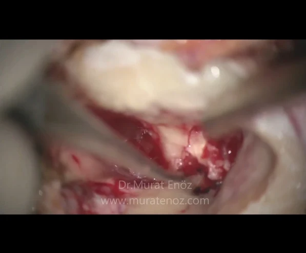
Foci of Inflammation Arising From Skin Cells In The Middle Ear and Mastoid Bone
What is a cholesteatoma?
A cholesteatoma is a rare abnormal collection of skin cells inside your ear in the middle ear and mastoid bone. It may have locally aggressive features such as tumoral tissue, and have properties that can damage the surrounding bone tissue and structures such as the surrounding cerebral cortex, brain, and facial nerve over time. It consists of trapped, reduced air contact, inflammatory ball-shaped skin cells. Cholesteatomas usually develop as cysts or bags that occur with the shedding of old skin layers accumulated in the middle ear. Over time, the cholesteatoma can increase in size, causing recurrent local infections, changes in the surrounding structure, and hearing loss by breaking down the delicate bones in the middle ear.
How does cholesteaoma occur?
It can be emphasized by two different mechanisms, congenital and acquired, in the development of choalesteatoma and the emergence of the inflammation focus with these locally aggressive features. Congenital cholesteatoma is a congenital cholesteatoma focus behind an intact eardrum. It is a condition in which a cholesteatoma occurs as a result of abnormal development of the structures inside the ear, but this is rare. In acquired cholesteatoma, it is explained as epithelial cells reaching the middle ear from the external ear canal or eardrum and forming the cholesteatoma focus there. If the eardrum collapses due to unequal pressure on a part of the eardrum, it may develop due to the migration of epithelial cells towards the middle ear. Cholesteatoma usually occurs due to poor Eustachian tube function and infection in the middle ear. It occurs when the Eustachian tube does not work properly. This is a thin tube that runs from the middle ear to the back of the nose. Its main function is to help maintain normal air pressure in the ear. Dead skin cells are normally expelled from the ear, but if the eardrum collapses, a pocket may appear towards the middle ear where dead skin cells can be collected. Cholesteatoma can occur after the eardrum has been damaged by an injury or infection, or after any type of ear surgery. In other words, a path is opened for the movement of epithelial cells from the external ear canal to the middle ear.
Cholesteaoma symptoms
Usually, cholesteatoma is found in only one ear and associated symptoms are in one ear. Symptoms and findings that may occur due to cholesteatoma:
• Persistent, often foul-smelling inflammatory discharge from the affected ear.
Generally, there is a relationship between water incontinence and the onset of ear discharge in patients with simple tympanic membrane perforation. In patients with cholesteaoma, middle ear infection and ear discharge may start unrelated to water contact, and the discharge may not decrease during drug treatment. Many patients have complaints of ear discharge that may run down their pillow during sleep, and that the discharge is very foul-smelling when it comes close to their nose. The prominent symptom of a cholesteatoma is persistent or frequently recurring painless otorrhea (ear discharge, often foul-smelling inflammatory discharge).
• Gradual hearing loss in the affected ear
Destruction of the middle ear tissues due to cholesteatoma, ossicular chain destruction and conductive hearing loss due to this, and sensorineural hearing loss due to recurrent infections are also present. Rarely, cholesteatoma creates a contact area between the fused ossicles, clearance of the cholesteatoma may cause an increase in conductive hearing loss.
• Dizziness
It is relatively rare. It is more likely to occur after repeated infections.
• Drainage and granulation tissue in the ear canal and middle ear
Depending on the chronic inflammatory discharge from the external ear canal, redness and dermatitis may occur on the skin in these areas. It may be helpful to use local cortisone lotions. In middle ear infections due to cholesteatoma, there may be no response to oral and local antibiotic treatment. Does not respond to antimicrobial therapy
• Tinnitus (tinnitus)
After recurrent suppurative otitis media attacks, destruction of inner ear cells and tinnitus may occur. Sometimes used local anti-iodine drops can also cause increased sensorineural hearing loss in the inner ear and tinnitus.
• Headache
In patients with cholesteatoma, it may be a precursor to complications such as meningitis, mastoiditis, or local infection.
• Feeling of "fullness" in one ear
Cholesteatoma and infection products, which usually fill the middle ear and mastoid cells, can cause this sensation.
• Weakness in half of your face
Paralysis of the feet or asymmetric decrease in facial movements may be a sign of facial paralysis complication, and surgery to remove cholesteatoma may be required in early edema.
Sometimes cholesteatoma initially presents with signs of intracranial complications, including:
• Sigmoid sinus thrombosis
• Epidural abscesses
• Meningitis
Some may experience some mild discomfort or a feeling of fullness in their ear.
Cholesteaoma Diagnosis
Laboratory investigations or incisional biopsies are generally not necessary in the diagnosis of cholesteatomas because the diagnosis can be made based on physical examination and radiological findings. The diagnosis can be made after an otoscopic examination, which is performed in the presence of the typical history of patients with "smelly ear discharge that starts from time to time without water in the ear and resistant to medical treatment", and sometimes in the presence of complications such as meningitis, facial paralysis, mastoiditis, neck or brain abscess due to cholesteatomas.
Computed tomography (CT) scanning is the diagnostic method of choice for this cholesteatoma because of its ability to detect fine bone defects.
Histologically, surgically removed cholesteatoma specimens contain typical squamous epithelium. Histologically, it is indistinguishable from histology of sebaceous cysts or keratomas from any part of the body.
Audiometry should be performed prior to surgery whenever possible. Air and bone conduction, speech perception threshold, and speech discrimination score should be determined within a few weeks of the proposed surgical procedure.
Magnetic resonance imaging (MRI) is used when very specific problems are suspected, such as:
• Dural involvement or invasion
• Subdural or epidural abscesses
• Herniation Brain into the mastoid space
• Inflammation of the membranous labyrinth or facial nerve
• Sigmoid sinus thrombosis
• Meningitis
Types of Cholesteaoma
In general, the following 3 types of cholesteatoma are defined:
• Congenital cholesteatoma
• Primary acquired cholesteatoma
• Secondary acquired cholesteatoma
Unlike other cholesteatomas, the congenital type is normally defined behind an intact, normal-appearing tympanic membrane.
Treatment of Cholesteatoma
In the treatment of cholesteatoma, mastoidectomy operations are generally performed to clean the trapped areas of inflammation and to ventilate the middle ear. Generally, the procedure is started behind the ear or by making an incision. In addition to removing dead skin cells, removal of the sponge-like mastoid bone (part of the skull behind the ear) and repairing any holes in the eardrum can be done at the same time. In most cases, surgery is performed under general anaesthesia. The primary goal of the surgery is to remove the cholesteatoma to eliminate the infection and create a dry ear (eliminating the serious health risks associated with cholesteatoma). The second goal of surgery is both to remove the cholesteatoma and to improve hearing by reconstruction of the damaged middle ear ossicles and eardrum. In other words, while the primary goal is to protect the patient from cholesteatoma and related risks; The secondary aim is to protect or normalize the hearing levels of the patients.
Health problems and risks that may arise due to untreated or delayed cholesteaoma
In the photograph above, the soft tissue density filling the right middle ear and partial erosion of the middle ear ossicles are seen. It is seen that the ossicles are surrounded by soft tissue and cholesteatoma tissue. In tomography, you can compare the right and left middle ear cavity. The photo of the endoscopic ear examination performed due to the intermittent malodorous inflammatory discharge from the ear of the same patient is the first cover photo of the video below. In the photograph, granulation tissue originating from the middle ear and protruding from the hole in the eardrum in the attic region is seen.
Left untreated, cholesteaoma can cause damage to delicate structures near the middle of the middle ear, such as the tiny bones and organs needed for hearing and balance.
The risks associated with cholesteatoma can be summarized as follows:
• an ear infection that causes ear infection
• hearing loss that may be permanent
• dizziness
• tinnitus
• damage to the facial nerve, weakness in half of the face
Rarely, an infection can spread to the inner ear and brain and cause a brain abscess or meningitis.
A cholesteatoma consists of squamous epithelium that is trapped in the skull base and can erode and destroy important structures within the temporal bone. The potential to cause central nervous system (CNS) complications (eg, brain abscess, meningitis) makes it a potentially dangerous lesion. In general, a long-term period is required for these complications.
Frequently asked questions and answers about cholesteatoma
Cholesteatoma surgery: For the treatment of cholesteatoma, an operation called mastoidectomy is performed to remove the inflamed areas of the mastoid bone with air pores behind the auricle.
Cholesteatoma treatment: This subject has been summarized in detail above.
What are the symptoms of cholesteatoma?: This topic has been summarized in detail above.
Does cholesteatoma recur? : In patients who are poorly treated and the trapped inflammatory areas are not fully opened, cholesteatoma may re-grow in the bone.
Is a cholesteatoma a tumor? : A cholesteatoma is a ball of active inflammation that behaves like a tumor and destroys adjacent bone tissues.
How long does cholesteatoma surgery take? : Cholesteatoma surgery usually takes between 2-4 hours. Concomitant eardrum surgery and middle ear ossicles repair surgery affect the duration.
How can I make an appointment for cholesteatoma treatment and mastoidectomy operation in Istanbul?: You can send a WhatsApp message to Dr. Murat Enöz when you click on the WhatsApp icon in Column at the top and left of this website. It will be useful to bring the results of the previous hearing tests and ear tomography with you.
How many days off should I take for cholesteatoma treatment and mastoidectomy operation in Istanbul?: You must come for the coronavirus pcr test 1 day before the operation. On the day of the operation, you will stay in the hospital for 1 night. You will be discharged later, and it is ideal that you stop for at least 7-9 days afterwards. If you stay longer (2 weeks is more suitable).
Links to find detailed information: http://www.healthzoneturkey.xyz/2017/08/cholesteatoma-symptoms-diagnosis-treatment.html
Link group where you can find articles about Cholesteatoma published on this website >> https://www.ent-istanbul.com/search?q=Cholesteatoma
Murat Enoz, MD, Otorhinolaryngology, Head and Neck Surgeon - ENT Doctor in Istanbul
Private Office:
Address: İncirli Cad. No:41, Kat:4 (Dilek Patisserie Building), Postal code: 34147, Bakırköy - İstanbul
Appointment Phone: +90 212 561 00 52
Fax: +90 212 542 74 47


Comments
Post a Comment