Porminent Ear Surgery - Otoplasty
This is a aesthetic surgical procedure which seeks to correct the appearance of protruding ears. In case the auricle is turned outward, there are different otoplasty techniques that involve bringing the auricle closer to the skull. Apart from the otoplasty operation, there may be patients who want the prominent ear to be shaped. In order to avoid a misunderstanding here, I would like to emphasize that auto plastic operations are usually performed to change the angle of the auricle and send it backwards when viewed from the front. Reshaping the unique folds in the auricle is not easy. Auricular reduction operation, namely auricular reduction, can be performed as a separate procedure, but shaping the auricle is usually a very difficult and complicated operation. Otoplasty operations, which can be performed in office conditions and involve many different techniques from simple to complex, can be defined as reducing the angle between the auricle and the skull and moving the auricle backwards. Except for simple suture techniques; there are more complex and longer-lasting procedures that involve reshaping the cartilages and recreating the most dangerous angle. These operations can be performed under local anesthesia and in office conditions as well as in hospital conditions.
Names that can be used synonymously with Otoplasty operation:
- Pinnaplasty
- Ear Plastic Surgery
- Cosmetic Ear Surgery
- Ear Reshaping
- Auriculoplasty
- Surgical Correction of Prominent Ears
- Protruding Ear Surgery
You can find the details of the techniques related to prominent ear operation below.
Pinna (or Auricle) Anatomy
The external ear (pinna / auricle) of living things in nature, unlike the others, plays a small role in human hearing function.
You can find the names of anatomical points on pinna at the video above. Among the above anatomical points, insufficient antihelical fold angle (antihelix / anthelix anti-helix angle) and larger than normal conchal cartilage are the main problems causing prominent ear deformity.
Definition of Protruding Ear or Prominent Ear
Prominent ear deformity is defined as a be angled outward and obvious meaning earlap deformity. The most common congenital anomalies in the skull and facial area , although there is no consensus on a clear definition in the literature.
For the emergence of prominent ear deformity in one or more of the following anatomical deformities should be existed:
- Antihelical angle is insufficient. Without sufficient amount of antihelikal fold can cause the pinna position as angled outward.
- Conchal cartilage is more than normal. In this way, turning right out of the auricular conchal will be more apparent from the pit .
- Conchomastoid angle is more than the normal. In this way, even if structurally normal auricle will be directed outwardly bucket may become evident.
- Further development of earlap (rare).
Conchal protrusion together with lack of antihelical angle are coexisted in 90% of patients. All of these deformities are mostly genetic. Sometimes it can be found with hereditary diseases.
Protruding Ear Surgery - Mustardé Technique - Animated Presentation
The most well-known suture technique between prominent ear correction surgeries is Mustardé matrix suture technique.Description of animated pictures:
- The injector tip dipped in the creation of a space for the marking of the planned new antihelical angle
- Incision is made behind the pinna, with continued dissection appears to make the back side of the conchal cartilage (At this stage, further processing can be done in the form of removal of the ellipsoid skin island).
- Matrix sutures are placed in the marked area shown in the injector holes
- Skin and subcutaneous sutures are placed and the operation is terminated.
Suture technique otoplasty complication - chronic suture reactions
The suture materials used in this method are usually permanent and non-absorbable suture materials. When there is a reaction due to these suture materials, it can move from under the skin to the skin and when the sutures come out, it can cause recurrent chronic local abscesses and chronic inflammation. Some patients may prefer to wait like this instead of removing these stitches by applying antibiotic ointment behind the ear for years. If the sutures are cut and removed, there is a risk of reinstatement of the cartilages. Link to the article where you can find the behind-the-ear images of a patient who had previously undergone otoplasty with this technique in a different clinic >> Stitch Reaction Risks or Infection After Suture Technique Otoplasty!
This technique performed especially in excess conchal cartilage, in order to reduce the depth of the conchal bowl and approximate the auricle to the skull.
Description of animated pictures:
- A double-edged ellipsoid shape additional incision is made on the back of conchal cartilage.
- On the outside of the new cartilage incision, skin is dissected from cartilage on the anterior part elipsoid cartilage area.
- While ellipsoid cartilage islet become on the anterior, mutual slip provided of surrounding cartilages and sutured.
- Skin and subcutaneous sutures placed and operation is terminated.
- Incision is made behind the pinna, with continued dissection appears to make the back side of the conchal cartilage.
- Re-created in the fold between the fields and the incisions are made on the cartilage incision islands separated from the front portion of the skin is dissected edge regions. Using absorbable suture materials are sewn to create folds backwards. In fact, in the form of islands of cartilage ellipsoid can also be created separately. In this technique defined to obtain a natural angle. Permanent suture is not required.
Apart from that, the cartilage weaken or different techniques are available for the cartilage abrasion.
To get the best results, one or a few of these techniques can be applied to each patient by the surgeon.
Antibiotics and pain medication are usually given for a period of first one week.
Varying amounts are normal to see the following which depending on the size of the surgery and different modifications of techniques:
Source Links:
Prominent Ear - eMedicine World Medical Library - Medscape
Prominent Ears — plastic - School of Medicine
The correction of the prominent ear - Springer
AAFPRS - Otoplasty | Cosmetic Ear Surgery
Defining the protruding ear
Ear Surgery - American Society of Plastic Surgeons
Ear Reshaping - NHS Choices
Otoplasty - MayoClinic.com
Protruding Ear Surgery - Pitanguy Technique - Animated Presentation
This technique performed especially in excess conchal cartilage, in order to reduce the depth of the conchal bowl and approximate the auricle to the skull.
Description of animated pictures:
- A double-edged ellipsoid shape additional incision is made on the back of conchal cartilage.
- On the outside of the new cartilage incision, skin is dissected from cartilage on the anterior part elipsoid cartilage area.
- While ellipsoid cartilage islet become on the anterior, mutual slip provided of surrounding cartilages and sutured.
- Skin and subcutaneous sutures placed and operation is terminated.
Protruding Ear Surgery - Modified Technique
Different techniques that can be modified and defined essentially for each ear. Here is a technique that I wanted to share my favorite.
Description of modified technique:
- Incision is made behind the pinna, with continued dissection appears to make the back side of the conchal cartilage.
- Re-created in the fold between the fields and the incisions are made on the cartilage incision islands separated from the front portion of the skin is dissected edge regions. Using absorbable suture materials are sewn to create folds backwards. In fact, in the form of islands of cartilage ellipsoid can also be created separately. In this technique defined to obtain a natural angle. Permanent suture is not required.
Protruding Ear Surgery - Conchamastoid Technique
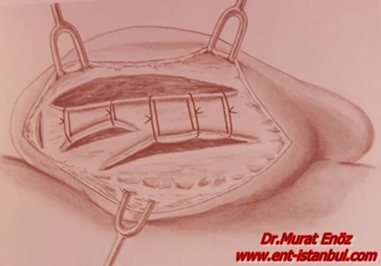
This technique is defined by the Furnas. The incision is made behind the pinna, mastoid dissection continued and the conchal cartilage and the back side is made visible. Permanent sutures placed
between conchal cartilage with periosteum which located on the mastoid bone behind the ear, pinna is pulled to back and inside side.
Apart from that, the cartilage weaken or different techniques are available for the cartilage abrasion.
To get the best results, one or a few of these techniques can be applied to each patient by the surgeon.
Postoperative Patient Care After Protruding Ear Surgery
Prominent ear bandage is applied for first 1 week after surgery and using rubber bands, bandanas or a tennis player which give less pressure for the next three weeks is recommended.
Antibiotics and pain medication are usually given for a period of first one week.
Varying amounts are normal to see the following which depending on the size of the surgery and different modifications of techniques:
- Pain in the ears and feeling the pressure to be due to the bandage.
- Edema and erythema on surgical area (usually seen in small amounts) .
- Incision scar behind the ear auricle can be visibl but usually it is unnoticed by people.
- Numbness around the incision behind the ear (this situation may improve in the months). Plaese remember that, shaping and hardening of the tissue after operation occur in a period of 2 months and for the full results and permanent harden up to 12 months . Therefore after surgery, ears plaster wrap or bandage application are used.
Bandana, Headband, Thin Hat For Pressuring on Ear
You can buy products that can be bought from a sports magazine in Istanbul and used for pressure on the ears. the use of these products may reduce the possibility of trauma to the ears; At the same time, it can prevent shape change by applying pressure. After about 2 months, the healing of the ear cartilage is mostly completed and we can end this pressure.
Cost of Otoplasty Operation in Istanbul
I usually use a modified technique (it takes a little longer and I don't use permanent sewing materials). When otoplasty operation is performed on average two ears, the price varies between 2500 - 3500 US Dollars according to hospitals. Processing fees may increase in luxury hospitals.
Why is a pressure bandage important after prominent ear surgery?
After prominent ear operations, there is a risk that the ears may return to their former shape as partially, with the cartilage memory. Those who have had prominent ear surgery using only the thread and suture method also have a higher risk of the ear becoming prominent again if these threads are dislodged or if the pressure is insufficient. I apply the modified technique prominent ear surgery and turbinate mastoid stitch technique more because of the high risk of reaction due to nonabsorbable suture materials and the risk of loosening of these suture materials and returning the auricle to its original shape in case of removal. Another benefit of the pressure ear bandage is that it prevents the formation of hematoma and the formation of the third space in the early postoperative period and reduces the risk of bleeding. I usually recommend keeping the tight ear wrap in place for about 1 week, then we can recommend our patients materials such as a tight bandana and a tight hat that are used by athletes and that tightly wrap the ears.
Before and After Photos for Ear Plastic Surgery in Istanbul, Turkey
On the following photographs you can find photos of patients who have undergone prominent ear aesthetic surgery using techniques of "Modified Technique" and "Concha-mastoid Technique"
Ear Lobe Reduction
This operation can be done together with prominent ear operation. It is a simple procedure that can only be done in office conditions and under local anesthesia. Although it is usually done in the form of suturing the incision sites end to end after the tissue in the form of a triangle is removed; There are also ear lobe reduction techniques, in which half-moon tissue is removed only from the lower part of the earlobe or by applying a different fel.
Ear Lobe Reduction Operation Cost in Istanbul
When this procedure is performed under office conditions and local anesthesia, it costs 500 USD for one ear and 800-1000 USD for both ears. If it is performed under hospital conditions, the total procedure fee is around 1500 USD for both ears. Our patients generally prefer to have the procedure performed under office conditions.
Care Recommendations After Ear Lobe Reduction Operation
After the procedures for the earlobe, if the prominent ear operation was not performed and only the earlobe reduction operation was performed; pressure dressing is not required. It is sufficient only to apply antibiotic ointment to the incision site and to care for the wound. Since we use suture materials that can dissolve in the body, all sutures dissolve spontaneously within 2-3 weeks.
The link group you can click to read the previously prepared and published articles about ear plastic surgery on this website >> https://www.ent-istanbul.com/search?q=ear+plastic+surgery
Source Links:
Murat Enoz, MD, Otorhinolaryngology, Head and Neck Surgeon - ENT Doctor in Istanbul
Private Office:
Address: İncirli Cad. No:41, Kat:4 (Dilek Patisserie Building), Postal code: 34147, Bakırköy - İstanbul
Appointment Phone: +90 212 561 00 52
Appointment Phone: +90 212 561 00 52
Fax: +90 212 542 74 47


.JPG)
.JPG)
.JPG)
.JPG)
.jpg)
.jpg)
.jpg)
.jpg)


.jpg)
.jpg)


Comments
Post a Comment