Triple-Layered Closure of an Oroantral Fistula

The nasal septum is the structure that divides the nasal cavity into two vertically and consists of the mucosa that covers the bone and cartilage. This structure allows each nasal cavity to work separately from each other. When the nasal septum is damaged due to various reasons, the resulting hole is called nasal septum perforation, septal hole, perforated nose septum. In this case, the location of the hole (front or back), the size of the hole (large or small) affect the amount of symptoms that will occur in patients. While almost no symptoms occur in small holes on the back of the septum; Dryness, bleeding, crusting, recurrent sinus infections and chronic atrophic mucosal changes may occur in the large holes in the anterior part of the septum.
Septum perforations may occur due to many different reasons such as surgical trauma, mechanical trauma, various drug use and cocaine use. You can find many articles on this subject at the link above.
Patient's History, Physical Examination and Surgical Technique
I would like to present a patient of yours who underwent septum perforation repair using temporal fascia, septal cartilage, and temporal fascia.
The patient's history and examination findings: The patient, who had undergone a tip plasty operation in a different clinic about 1 year ago and worked as a welding worker, applied to our office due to the sensation of dryness and burning in the nose that had occurred in the last 6 months. In the examination of the patient, it was observed that there was widespread metal dust in the nose, and the nasal mucosa was crusty and dry. A few mm diameter perforation was detected near the anterior part of the septum. The patient was advised to use nasal moisturizing products, antibiotic eye ointment, saline nasal irrigation, and the use of a protective mask during welding was recommended. Due to the increase in the patient's complaints within 6 months and the enlargement of the hole in the nasal septum, the operation for closure of the septum perforation was planned for the patient.
Surgical planning: Normally, there are many surgical techniques defined for closure of septum perforation. There are simple local mucosal flap techniques, techniques using cartilage grafts, techniques using temporal fascia, perichondrium and various artificial membranes, techniques using vomer and ethmoid perpendicular plate. Since the patient I shared with you had to come into contact with the smoke of a hot and dry welding machine due to his profession (he states that he does not always use a protective mask even though it is recommended to use a protective mask), the nasal mucosa during the examination is quite dry and unhealthy, we planned a repair technique that includes 3 layers of tissue.
Preoperative preparation: Nasal irrigation with saline and rifampicin for 1 week was recommended to the patient preoperatively. The patient was advised to consume fresh vegetables and fruits with high water content, and not to consume strong black tea and coffee by mouth.
Surgical technique: The operation was planned under general anesthesia. After local anesthetic infiltration to the right temple area and the perforation edges of the nose, an approximately 2.5 cm horizontal incision was made in the right temple area. A 2x3 cm graft was taken from the temporalis muscle fascia. Local mucosal flaps (upper and lower flaps) were prepared from the inside of the nose (with the closed technique) from the edges of the perforation and tilted towards the perforation area. A 1x1.5 cm cartilage graft was placed on the right side and 2x1 cm temporalis fascia on it and 1x1 cm temporalis fascia on the left side. These tissues were sutured in full thickness with 4/0 vicryl. Below you can find the photographs taken from the inside of the patient's right and left nostrils. After the procedure was completed, a pressure dressing was placed on the head and internal nasal silicone splints were placed in the nose. The operation was terminated.

Temporalis Fascia Grafting:
As seen in the photo on the right, a graft was obtained from the fascia of the temporal muscle after the incision made in the right temporal region. Intracutaneous sutures were made using self-absorbable suture material on the incision area on the skin of the line and 3 additional incision tightening sutures were placed. The printed head wrap was removed after 1 day, leaving the wound open.
Postoperative care recommendations: Nasal irrigation with a mixture of rifampicin + saline, local eye ointment application and oral antibiotic tablets were recommended to the patient. It was recommended that silicone splints remain in the nose for longer than 1 month.
 |
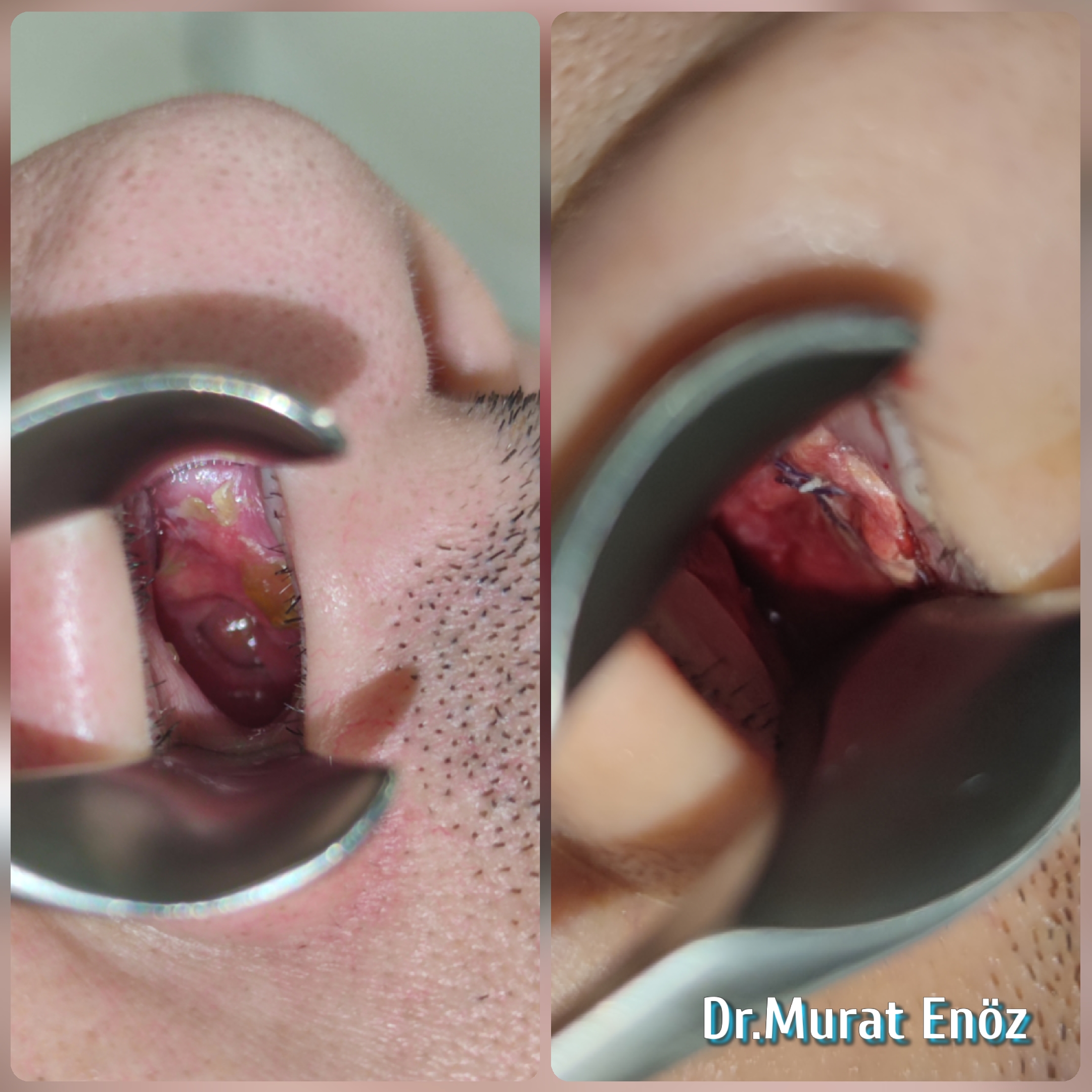
| Photograph taken from the right nostril, pre-operative and post-operative image. There is a dark purple appearance of the temporalis fascia graft and 4/0 vicryl suture material covering the perforation area (not visible because the cartilage graft is under the fascia). |
 |
| Photograph taken from the right nostril, pre-operative and post-operative image. There is a dark purple appearance of the temporalis fascia graft and 4/0 vicryl suture material covering the perforation area (not visible because the cartilage graft is under the fascia). |
 |
| Photo taken from the left nostril, before and after the operation. There is a dark purple appearance of the temporalis fascia graft (more packed and inserted into the hole) and 4/0 vicryl suture material covering the perforation area. |
 |
| Photo taken from the left nostril, before and after the operation. There is a dark purple appearance of the temporalis fascia graft (more packed and inserted into the hole) and 4/0 vicryl suture material covering the perforation area. |
Suggested link >> Septal Perforation - StatPearls - NCBI Bookshelf
Murat Enoz, MD, Otorhinolaryngology, Head and Neck Surgeon
Private Office:
Address: İncirli Cad. No:41, Kat:4 (Dilek Patisserie Building), Postal code: 34147, Bakırköy - İstanbul
Appointment Phone: +90 212 561 00 52
Appointment Phone: +90 212 561 00 52
Fax: +90 212 542 74 47

Comments
Post a Comment